
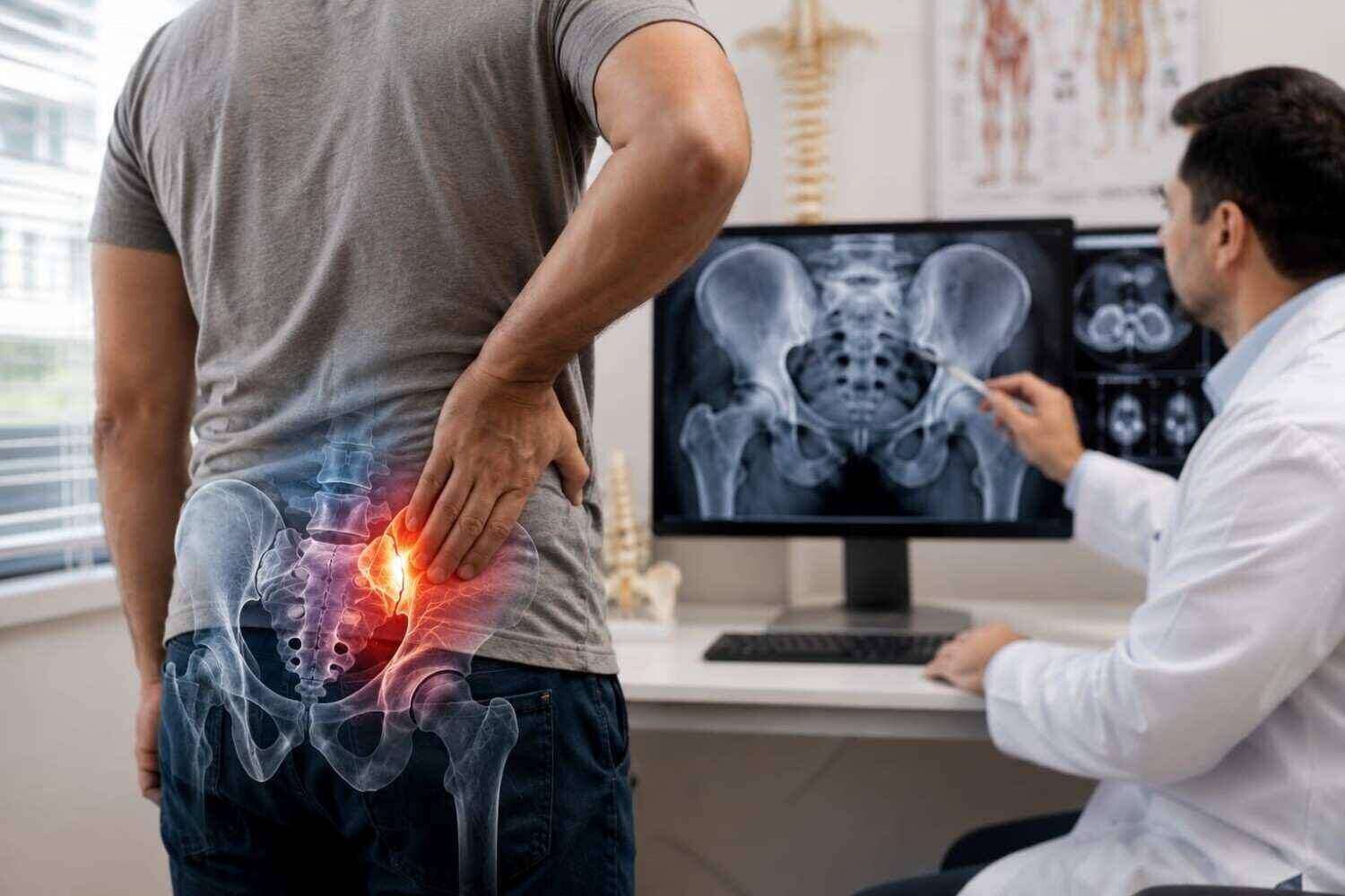
SI Joint Pain Treatment in Laredo
If you've been told your back pain "isn't coming from your spine," sacroiliac joint dysfunction may be the answer you've been searching for. Research suggests this single joint is responsible for 15% to 30% of chronic lower back pain cases — yet it remains one of the most frequently missed diagnoses in pain medicine. Here's what the journey from first symptoms to effective SI joint pain treatment in Laredo actually looks like.
What Is the Sacroiliac Joint — and Why Does It Cause So Much Pain?
The sacroiliac (SI) joint connects the sacrum — the triangular bone at the base of your spine — to the iliac bones of the pelvis on either side. It's a weight-bearing joint that absorbs shock between your upper body and legs during every step you take. For patients in physically demanding occupations common across Webb County — logistics, warehousing, oil and gas, and ranching — that's a joint under constant mechanical stress.
What makes SI joint pain particularly difficult to identify is where it shows up. The joint receives nerve supply from the ventral rami of L4–S2 and dorsal rami of L5–S2, which means pain signals can travel into the gluteal region, lower back, groin, and even down the leg — mimicking sciatica or lumbar disc disease. As Vleeming et al. note in the Journal of Anatomy, "a thorough understanding of SI joint neurology and biomechanics is essential for interpreting diagnostic injections and directing targeted therapy."
That overlap with sciatica symptoms is exactly why so many patients spend months — sometimes years — receiving treatment for the wrong diagnosis. If you've been managing what feels like sciatica without lasting improvement, SI joint dysfunction deserves serious consideration.
Common Causes of SI Joint Dysfunction
The SI joint can become painful through several distinct pathways:
Traumatic injury — A fall onto the buttocks, motor vehicle accident, or sudden twisting force can disrupt the joint's ligamentous support. Auto accident patients in Laredo frequently present with SI joint involvement alongside lumbar and cervical complaints.
Pregnancy-related ligamentous laxity — Hormonal changes during pregnancy loosen the pelvic ligaments, which can destabilize the SI joint. Postpartum pain that lingers below the belt line is a classic presentation.
Degenerative arthritis — Like any synovial joint, the SI joint is subject to osteoarthritic change over time, particularly in patients with a history of heavy physical labor.
Inflammatory spondyloarthropathy — Conditions such as ankylosing spondylitis or psoriatic arthritis can target the SI joint specifically, producing inflammatory rather than mechanical pain.
Leg-length discrepancy — Even a modest difference in leg length alters pelvic mechanics and can create asymmetric loading across the SI joint over years.
Patients with hypermobility conditions — such as those discussed in our article on Ehlers-Danlos syndrome and joint stability — are also at elevated risk because their ligamentous laxity reduces the structural support the SI joint depends on.
It's also worth noting that excess body weight increases mechanical load on the pelvis. For patients managing weight alongside joint pain, sustainable weight loss strategies can meaningfully reduce the stress placed on the SI joint over time.
How SI Joint Dysfunction Is Diagnosed
This is where the patient journey most often stalls. Because SI joint pain overlaps with lumbar disc pathology, hip arthritis, and even inflammatory conditions like lupus, arriving at the correct diagnosis requires a structured, stepwise approach.
As Cohen SP notes in Anesthesia & Analgesia, "sacroiliac joint pain remains one of the most underdiagnosed causes of axial low back pain, necessitating a high index of clinical suspicion and confirmatory diagnostic blocks."
Step 1 — Clinical History and Pain Mapping
The typical SI joint pain pattern includes:
Pain located within 2–3 cm inferior and medial to the posterior superior iliac spine (PSIS)
Unilateral buttock or posterior hip pain that may radiate into the groin or thigh
Pain worsened by prolonged sitting, rising from a chair, climbing stairs, or lying on the affected side
No neurological deficits (numbness, weakness, or reflex changes) in most mechanical cases
Step 2 — Physical Examination Maneuvers
No single test confirms SI joint dysfunction, but a cluster of at least three positive provocation tests significantly increases diagnostic accuracy. The most validated maneuvers include:
Distraction test — Pressure applied to the anterior iliac crests to stress the anterior SI ligaments
Thigh thrust — Axial load through the femur with the hip flexed to 90 degrees
FABER test (Flexion, ABduction, External Rotation) — Assesses hip and SI joint mobility simultaneously
Compression test — Lateral pressure on the iliac crests to compress the SI joint
Step 3 — Image-Guided Diagnostic Injection
When clinical suspicion is high after examination, the gold standard for confirmation is a fluoroscopy-guided intra-articular anesthetic injection. If a patient reports 75% or greater pain relief following the injection, SI joint dysfunction is confirmed as the primary pain generator. This step is critical — it simultaneously rules out other pain sources and guides the treatment plan.
At Pain Management of Laredo, fluoroscopy-guided SI joint injections are performed with precision imaging to ensure accurate needle placement, which is essential both for diagnostic validity and therapeutic effect.
SI Joint Pain Treatment Options: A Comparison
Once the diagnosis is confirmed, treatment follows a stepwise progression from conservative to interventional. Results vary by patient, and your specialist will recommend a personalized approach based on symptom severity, duration, and response to each stage of care.
| Treatment | How It Works | Duration of Relief | Recovery Time | Best For |
|---|---|---|---|---|
| Physical Therapy / Pelvic Stabilization | Strengthens muscles supporting the SI joint; corrects movement patterns | Ongoing with compliance | None | Mild-to-moderate mechanical dysfunction |
| SI Joint Corticosteroid Injection | Fluoroscopy-guided injection of anti-inflammatory medication into the joint | 2–6 months (varies) | 24–48 hours of soreness | Confirmed SI joint inflammation or arthritis |
| Radiofrequency Ablation (RFA) | Thermal energy disrupts the nerve branches transmitting pain signals | 9–18 months typically | 1–2 weeks | Patients with confirmed SI joint pain who respond to diagnostic blocks |
| Minimally Invasive SI Joint Fusion | Triangular titanium implants stabilize the joint permanently | Long-term / durable | 2–6 weeks, activity-dependent | Severe, refractory SI dysfunction failing conservative and injection therapy |
Conservative and Physical Approaches
Pelvic stabilization therapy — including targeted exercises, SI joint bracing, and movement retraining — is the appropriate starting point for most patients. Yoga-based approaches can be integrated carefully; poses such as bridge with a block between the thighs, modified triangle, and supine figure-4 stretches may support SI joint stability when performed with proper alignment guidance.
Fluoroscopy-Guided Injections
For patients whose pain persists despite conservative care, image-guided SI joint injections deliver corticosteroid medication directly into the joint space. The fluoroscopic guidance is non-negotiable — the SI joint's irregular anatomy makes blind injections unreliable. Beyond pain relief, the injection response provides diagnostic clarity that informs all subsequent treatment decisions.
Radiofrequency Ablation
When injections provide meaningful but temporary relief, radiofrequency ablation (RFA) of the lateral branch nerves supplying the SI joint can extend that relief significantly. The procedure uses controlled heat to interrupt pain signal transmission and typically provides 9 to 18 months of benefit, with the option to repeat as nerves regenerate.
Minimally Invasive SI Joint Fusion
For patients with debilitating SI joint dysfunction who have not responded to conservative measures and multiple injections, minimally invasive fusion offers a durable surgical solution. The landmark INSITE randomized controlled trial demonstrated that fusion using triangular titanium implants produced statistically significant improvements in pain and disability at 24 months compared to continued conservative management. The INSITE trial investigators noted that "minimally invasive fusion represents a durable surgical solution for patients with debilitating SI joint dysfunction who have exhausted conservative measures."
Patients in Laredo who reach this stage can access referral pathways to South Texas orthopedic and spine specialists through coordinated care with Pain Management of Laredo.
What Laredo Patients Should Know Before Their First Appointment
Webb County residents often face a meaningful access gap when it comes to interventional pain care. Without a local specialist, the alternative is a multi-hour drive to San Antonio or Houston — a significant burden for anyone already in pain and managing work and family obligations.
A few practical points for patients preparing for their first SI joint evaluation:
Bring a pain diary. Document where your pain is located (a body diagram helps), what activities worsen it, and what provides temporary relief. Note whether the pain is worse on one side.
List all previous imaging. MRI and CT reports of the lumbar spine are useful for ruling out competing diagnoses, even if they came back "normal." A normal lumbar MRI in a patient with SI joint pain is actually a meaningful data point.
Be honest about activity demands. Patients in physically demanding jobs — driving trucks across the border, working warehouse shifts, managing ranching operations — have different biomechanical profiles than sedentary patients. This context shapes the treatment plan.
Ask about bilingual care. Many Laredo patients are more comfortable discussing pain symptoms in Spanish. Dr. Memon's practice serves the bilingual community, and communicating clearly about your pain is essential to accurate diagnosis.
For patients involved in auto accidents, it's also worth knowing that SI joint injuries from the impact forces of a collision are well-documented. The practice accepts letters of protection (LOP) for personal injury cases, so insurance or legal status should not be a barrier to getting evaluated.
Frequently Asked Questions
How do I know if my back pain is coming from the SI joint versus a disc or nerve?
The location is the first clue — SI joint pain typically sits below the belt line, often one-sided, and may radiate into the buttock or groin rather than shooting down the entire leg with numbness or tingling. However, the only reliable way to distinguish SI joint pain from lumbar disc or nerve pathology is through a structured physical examination followed by a diagnostic fluoroscopy-guided injection. Imaging alone (X-ray, MRI) is not sufficient to confirm SI joint dysfunction.
How long does an SI joint injection take, and what should I expect afterward?
The injection procedure itself typically takes 15 to 30 minutes, including positioning and fluoroscopic guidance. Most patients experience mild soreness at the injection site for 24 to 48 hours afterward. Pain relief — if the SI joint is the primary pain generator — usually begins within a few days as the corticosteroid takes effect. You should arrange for someone to drive you home following the procedure.
Is SI joint fusion a major surgery?
Minimally invasive SI joint fusion is performed through a small incision, typically under general or regional anesthesia, and most patients go home the same day or after one overnight stay. It is significantly less invasive than open spinal fusion procedures. Recovery involves protected weight-bearing for several weeks, with most patients returning to light activity within 2 to 6 weeks depending on their baseline health and occupational demands. Your specialist will outline a personalized recovery timeline.
Can SI joint pain come back after treatment?
Injections and radiofrequency ablation provide temporary relief — the duration varies by patient, but RFA typically lasts 9 to 18 months before nerves regenerate. Minimally invasive fusion is intended as a long-term solution, with the INSITE trial data supporting durable outcomes at 24 months. For patients managing underlying inflammatory conditions, ongoing disease management is also necessary to prevent recurrence.
Take the Next Step Toward SI Joint Relief in Laredo
You don't have to drive to San Antonio to get answers. At Pain Management of Laredo, Dr. Rehan Memon offers board-certified, interventional pain care — including fluoroscopy-guided diagnostic injections, corticosteroid treatment, and coordinated referral for minimally invasive fusion — right here in Webb County.
If you've been living with unexplained lower back or buttock pain, call us at +1 956-962-7173 or visit our office at 8511 McPherson Rd Ste. 204, Laredo, TX to schedule a consultation. Same-week appointments are available, and we proudly serve bilingual patients throughout Laredo and the surrounding South Texas region. For additional context on back pain research and treatment options, the National Institute of Arthritis and Musculoskeletal and Skin Diseases offers a reliable federal resource.
Medically reviewed by Dr. Rehan Memon, Board-certified pain specialist and anesthesiologist with 25+ years of experience
Interventional Pain Management Specialist at Pain Management of Laredo
Last reviewed: May 2026
This article is for informational purposes only and does not constitute medical advice. Please consult a qualified healthcare provider for diagnosis and treatment.